
Terapia farmacologica (testo con alcuni contenuti sponsorizzati)
Una volta eseguito un corretto iter diagnostico e verificata l’alterazione metabolica alla base della formazione dei calcoli, è possibile instaurare una corretta terapia medica. Grazie alla comprensione del meccanismo patologico alla base della formazione di calcoli (descritto nel capitolo “calcoli renali cause”), è possibile prescrivere programmi terapeutici basati su terapie selettive.
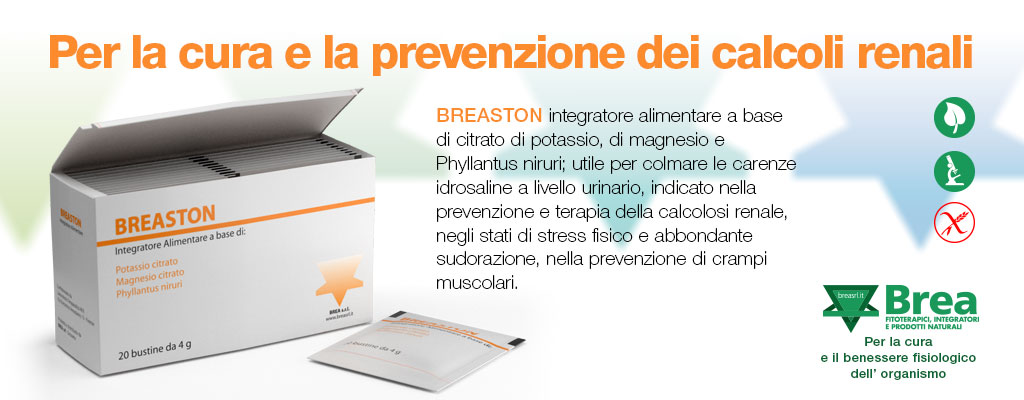
Banner pubblicitario
La terapia farmacologica della calcolosi si prefigge i seguenti obiettivi:
- curare la patologia metabolica di fondo
- inibire la formazione di nuovi calcoli
- curare le altre complicanze (non solo quelle renali) della patologia metabolica
- minimizzare i possibili effetti collaterali
I farmaci che possono essere impiegati nella terapia medica della calcolosi sono i seguenti:
- Diuretici tiazidici (idroclorotiazide, clortalidone, indapamide)
- Fosfato di cellulosa di sodio
- Ortofosfati
- Citrato di potassio
- Allopurinolo
- Gluconato di magnesio
- Piridossina (Vitamina B6)
- Captopril
- alfa mercapto propionil glicina
I tiazidici vengono impiegati nei casi di ipercalciuria da assorbimento intestinale perchè stimolano il riassorbimento di calcio da parte del rene. La prescrizione di tiazidici e di citrato di potassio, combinata con una restrizione di calcio e di ossalati con la dieta, è in grado sia di correggere la ipercalciuria che di limitare l’osteopenia solitamente associata a questa patologia
In caso di fallimento dei tiazidici, è possibile impiegare l’ortofosfato. Il fosfato di cellulosa di sodio è una resina a scambio ionico che lega il calcio inibendone l’assorbimento intestinale. Fu inizialmente impiegata nei casi di ipercalciuria da riassorbimento, ma il suo utilizzo è stato oggi abbandonato, nonostante gli entusiasmi iniziali, per la presenza di effetti collaterali.
I tiazidici in combinazione con il citrato di potassio sono la terapia di prima linea nella ipercalciuria renale. Nei casi in cui l’ipercalciuria è causata da un iperparatirodismo, la terapia non è medica ma chirurgica, e consiste nella rimozione dell’adenoma paratiroideo o dell’ intera ghiandola.
L’ iperuricosuria può essere corretta limitando l’assunzione di proteine animali e grazie alla prescrizione di allopurinolo e di citrato di potassio. L’ allopurinolo riduce la sintesi di acido urico, mentre il citrato di potassio ne aumenta la solubilità nelle urine.
L’iperossaluria intestinale viene corretta aumentando l’assunzione di liquidi e di calcio con la dieta. Il calcio, legandosi all’ ossalato nell’ intestino, ne riduce l’assorbimento. Nei casi in cui l’ iperossoaluria si associa ad una ipopotassemia, sarà necessario aggiungere citrato di potassio alla terapia.
L’ipocitraturia, a prescindere dalla causa, viene facilmente corretta con l’assunzione di citrato di potassio.
L’ ipomagnesuria viene curata aumentando l’assunzione di magnesio. Solitamente vengono utilizzati integratori a base di citrato, potassio e magnesio. Questo perché l’assunzione del solo magnesio causa diarrea e altri effetti collaterali intestinali.
Esistono scarse opzioni farmacologhe per la cistinuria. La terapia di questa alterazione metabolica si basa principalmente sull’ assunzione di abbondanti liquidi, alcalinizzazione delle urine e riduzione del sale nella dieta. In alcuni casi è possibile utilizzare l’alfa mercapto propionil glicina (nome commerciale Thiola), un farmaco che, legandosi alla cistina, ne favorisce l’escrezione. Tuttavia vi è una scarsa tolleranza a questo farmaco per i suoi effetti collaterali.
I calcoli infettivi a base di struvite vanno rimossi chirurgicamente. Non è raccomandabile la dissoluzione di questo tipi di calcoli tramite farmaci. Le infezioni recidivanti vanno curate con una opportuna terapia antibiotica e l’adozione delle misure preventive descritte altrove nel nostro sito (cistite, infezioni vie urinarie).
I calcoli vescicali devono essere rimossi tramite trattamento endoscopico. Per evitare la recidiva di questo tipo di calcoli è necessario risolvere la patologia ostruttiva che li ha causati (ipertrofia prostatica, stenosi uretrale).
ESWL, Terapia endoscopica e chirurgica (torna su)
Nei capitoli precedenti abbiamo descritto il trattamento conservativo e farmacologico dei calcoli. Tuttavia, se queste terapie iniziali non funzionano, come eliminare i calcoli renali?

L’obiettivo della terapia chirurgica è rimuovere la maggiore quantità possibile di calcoli in un unico intervento minimizzando le complicanze.
Le tecniche attive per eliminare i calcoli renali sono quattro, elencate in ordine di invasività:
- ESWL
- URS
- PCNL
- Laparoscopia (tradizionale o robotica)
Queste tecniche possono essere anche usate in combinazione.
La litotrissia extracorporea (ESWL), volgarmente nota come il “bombardamento per calcoli renali”, si esegue in regime ambulatoriale. Questa procedura, a differenza del trattamento chirurgico, non richiede il ricovero e viene indicata sia per il trattamento dei calcoli renali che di quelli ureterali.
Si avvale di un macchinario esterno che produce onde d’ urto, impulsi meccanici ad alta intensità. Si utilizzano per l’identificazione del calcolo ai reni varie tecniche radiologiche di puntamento (radioscopia e/o ecografia).
Dopo aver individuato il calcolo, l’operatore, che può essere sia un urologo che un tecnico abilitato, indirizza le onde d’urto direttamente sul calcolo per frantumarlo. I calcoli vengono ridotti in un materiale simile a sabbia, i cui i granelli possono essere espulsi attraverso l’urina. I calcoli renali di grandi dimensioni possono richiedere più sedute di trattamento.
Se un calcolo non viene espulso entro un mese, viene preso in considerazione un trattamento chirurgico eseguito in regime di ricovero. L’urologo può svolgere varie procedure per rompere e rimuovere i calcoli renali.
L’ureteroscopia (URS) si basa sull’ utilizzo di un ureteroscopio, uno strumento provvisto di fibre ottiche o di una telecamera digitale alla punta. L’uretroscopio viene introdotto prima nell’uretra, poi in vescica e di qui nell’uretere. Una volta individuato il calcolo, l’urologo lo rimuove mediante un piccolo cestello introdotto in un canale operativo dell’ureteroscopio o lo frantuma con un laser o con un dispositivo simile. I frammenti vengono successivamente eliminati dal paziente. L’ureteroscopia viene eseguita in anestesia generale o spinale.
La Nefrolitotomia percutanea (PCNL) è una procedura in cui i calcoli vengono rimossi attraverso una piccola incisione sul fianco del paziente. Questa procedura viene eseguita in anestesia locale e sedazione endovenosa. La rimozione percutanea (ossia attraverso la pelle) dei calcoli (litotomia) si esegue mediante un accesso direttamente sul rene attraverso il fianco. Un particolare ago ed un “filo guida” sono usati per guadagnare l’accesso all’interno delle vie escretrici renali.
L’incisione praticata viene dilatata e successivamente viene introdotto il nefroscopio, lo strumento attraverso il quale l’urologo frantuma il calcolo e lo estrae. Esistono vari modi con i quali si possono frantumare i calcoli sia in corso di URS che di PCNL, basati sull’ utilizzo di vari macchinari:
- La Litotrissia ultrasonicautilizza onde meccaniche ad alta frequenza (ultrasuoni) che vengono generate da una sonda elettrica che viene posta a contatto diretto con il calcolo. I frammenti prodotti dalla frantumazione del calcolo vengono eliminati dal paziente o sono rimossi chirurgicamente.
- La Litotrissia balisticaimpiega una sonda che genera vibrazioni meccaniche per rompere i calcoli, sfruttando un principio fisico analogo a quello del martello pneumatico. Si può usare sia in corso di ureteroscopia che di PCNL. Alcune sonde sono provviste di entrambi i dispositivi: ultrasonico e balistico.
- La Litotrissia elettroidraulica (EHL)impiega una sonda flessibile che produce onde d’ urto tramite l’elettricità. La sonda è posizionata vicino al calcolo attraverso un ureteroscopio flessibile. I frammenti possono essere eliminati dal paziente o essere estratti. L’EHL richiede l’anestesia generale e può essere utilizzata per rompere calcoli in qualsiasi distretto del sistema urinario.
- La litotrissia laser usa un laser ad Olmio per distruggere i calcoli. Anche questa tecnica si può usare tanta per la URS che per la PCNL.
Dopo l’esecuzione di una URS(ureteroscopia) o di una PCNL(Nefrolitotomia percutanea), il vostro urologo potrà decidere nella maggior parte dei casi di posizionare uno “stent a doppio J” nell’ uretere. Si tratta di un piccolo tubicino che consente il passaggio delle urine ed evita al paziente fastidiose coliche renali.
La chirurgia aperta viene praticata in anestesia generale. Si esegue un’incisione al fianco del paziente e il calcolo viene estratto attraverso un taglio dell’uretere o del rene. Per la maggior parte dei pazienti è necessario un ricovero prolungato e il recupero completo richiese parecchie settimane.
Cenni storici (torna su)
Prima dell’avvento dell’endourologia, i calcoli renali venivano rimossi solo ed esclusivamente tramite chirurgia a cielo aperto. Questo tipo di approccio è efficace ma è altresì gravato da un alto tasse di complicanze.
All’ inizio degli anni ’80 è stata introdotta la ESWL (litotrissia extracorporea con onde d’ urto): questa tecnica presenta una eccellente grado di sicurezza per il paziente ed un discreto tasso di successo. Nello stesso periodo è nata anche la PCNL (Nefrolitotomia percutanea) che oggigiorno viene considerata l’approccio di riferimento per i calcoli grandi e complessi.
Negli ultimi 20 anni, grazie al progresso della tecnologia, è aumentata anche la frequenza di utilizzo della URS (Ureterorenoscopia). Ancora più recentemente sono state utilizzate con successe anche le tecniche laparoscopiche e robotiche. Grazie a queste nuove tecnologie la frequenza di utilizzo della chirurgia open per i calcoli renali si è ridotta notevolmente passando dal 26% al 3%.
Indicazioni (torna su)
La decisione su quale tecnica utilizzare per eliminare i calcoli renali non è sempre semplice. La scelta dell’approccio dipende dal caso clinico, dall’ esperienza dell’urologo e dalla disponibilità dello strumentario a disposizione.
Per la programmazione della terapia chirurgica bisogna prendere in considerazione in primo luogo i fattori relativi al calcolo: grandezza, numero, localizzazione e composizione. La maggior parte dei calcoli renali ha dimensioni inferiori al centimetro ed è asintomatica per il 50-60% dei casi. Tuttavia, con il passare del tempo, un calcolo può ingrandirsi e dare problemi.
L’ESWL, volgarmente nota come bombardamento, è considerata la terapia di prima scelta per i calcoli di piccole dimensioni. Il bombardamento è infatti la metodica meno invasiva, è in grado di ottenere una buona percentuale di successo nell’eliminazione del calcolo (compresa tra il 50% ed il 90%) e non richiede una grande curva di apprendimento per l’urologo.
Negli ultimi tempi, in alternativa all’ESWL, si sta diffondendo anche l’URS con ureteroscopio flessibile per i calcoli di dimensioni inferiori al centimetro. Questa metodica, pur richiedendo il ricovero in regime ospedaliero e l’anestesia, possiede, se condotta da urologi esperti, percentuali di successo comprese tra l’80% ed il 90% (superiori a quelle dell’ESWL).
I calcoli di medie dimensioni (tra gli 1 ed i 2 centimetri), possono essere trattati con il bombardamento, l’URS e/o la PCNL. Se il calcolo non è localizzato nel calice inferiore, l’ESWL è la terapia di prima linea.
La PCNL è il trattamento di prima scelta per i calcoli di grandi dimensioni (superiori ai 2 cm) e per i calcoli di medie dimensioni localizzati nel calice inferiore. Il tasso di successo della PCNL è relativamente indipendente dalla dimensione e dalla composizione del calcolo. Tuttavia la PCNL ha un tasso di complicanze compreso tra il 20 ed il 30%.
Altro fattore da considerare è la composizione del calcolo. I calcoli di cistina, di fosfato di calcio (specialmente quelli di brushite) e quelli di ossalato di calcio monoidrato sono i più resistenti al bombardamento. In particolare, calcoli di estrema durezza sono difficili da frantumare tramite le onde d’ urto generate dal bombardamento.
Informazione presuntive sulla durezza e la consistenza di un calcolo possono essere dedotte dalla TAC, ed in particolare dalla HU, l’unità di misura della massa. Altro fattore che limita l’efficacia dell’ESWL è la distanza tra la cute ed il calcolo, condizione che si verifica ad esempio in un paziente obeso.


BIBLIOGRAFIA
- Aboumarzouk, O.M., et al. Flexible ureteroscopy and laser lithotripsy for stones >2 cm: a systematic review and meta-analysis. J Endourol, 2012. 26: 1257.
- Akman, T., et al. Comparison of percutaneous nephrolithotomy and retrograde flexible nephrolithotripsy for the management of 2-4 cm stones: a matched-pair analysis. BJU Int, 2012. 109: 1384.
- Argyropoulos, A.N., et al. Evaluation of outcome following lithotripsy. Curr Opin Urol, 2010. 20: 154.
- Assimos, D.G., et al. The role of open stone surgery since extracorporeal shock wave lithotripsy. J Urol, 1989. 142: 263.
- Auer, B.L., et al. The effect of ascorbic acid ingestion on the biochemical and physicochemical risk factors associated with calcium oxalate kidney stone formation. Clin Chem Lab Med, 1998. 36: 143.
- Auge, B.K., et al. Ureteroscopic management of lower-pole renal calculi: technique of calculus displacement. J Endourol, 2001. 15: 835.
- Barcelo, P., et al. Randomized double-blind study of potassium citrate in idiopathic hypocitraturic calcium nephrolithiasis. J Urol, 1993. 150: 1761.
- Bas, O., et al. Management of calyceal diverticular calculi: a comparison of percutaneous nephrolithotomy and flexible ureterorenoscopy. Urolithiasis, 2015. 43: 155.
- Basiri, A., et al. Comparison of safety and efficacy of laparoscopic pyelolithotomy versus percutaneous nephrolithotomy in patients with renal pelvic stones: a randomized clinical trial. Urol J, 2014. 11: 1932.
- Becker, G. Uric acid stones. Nephrology, 2007. 12: S21.
- Beltrami, P., et al. The endourological treatment of renal matrix stones. Urol Int, 2014. 93: 394.
- Bichler, K.H., et al. Indications for open stone removal of urinary calculi. Urol Int, 1997. 59: 102.
- Bichler, K.H., et al. Urinary infection stones. Int J Antimicrob Agents, 2002. 19: 488.
- Binbay, M., et al. Evaluation of pneumatic versus holmium:YAG laser lithotripsy for impacted ureteral stones. Int Urol Nephrol, 2011. 43: 989.
- Biyani, C.S., et al. Cystinuria—diagnosis and management. EAU-EBU Update Series 2006. 4: 175.
- Borghi, L., et al. Comparison of two diets for the prevention of recurrent stones in idiopathic hypercalciuria. N Engl J Med, 2002. 346: 77.
- Borghi, L., et al. Nifedipine and methylprednisolone in facilitating ureteral stone passage: a randomized, double-blind, placebo-controlled study. J Urol, 1994. 152: 1095.
- Borghi, L., et al. Randomized prospective study of a nonthiazide diuretic, indapamide, in preventing calcium stone recurrences. J Cardiovasc Pharmacol, 1993. 22 Suppl 6: S78.
- Borghi, L., et al. Urinary volume, water and recurrences in idiopathic calcium nephrolithiasis: a 5-year randomized prospective study. J Urol, 1996. 155: 839.
- Brandt, B., et al. Painful caliceal calculi. The treatment of small nonobstructing caliceal calculi in patients with symptoms. Scand J Urol Nephrol, 1993. 27: 75.
- Brocks, P., et al. Do thiazides prevent recurrent idiopathic renal calcium stones? Lancet, 1981. 2: 124.
- Burgher, A., et al. Progression of nephrolithiasis: long-term outcomes with observation of asymptomatic calculi. J Endourol, 2004. 18: 534.
- Cameron, M.A., et al. Uric acid nephrolithiasis. Urol Clin North Am, 2007. 34: 335.
- Campschroer, T., et al. Alpha-blockers as medical expulsive therapy for ureteral stones. Cochrane Database Syst Rev, 2014. 4: CD008509.
- Chan, L.H., et al. Primary SWL Is an Efficient and Cost-Effective Treatment for Lower Pole Renal Stones Between 10 and 20 mm in Size: A Large Single Center Study. J Endourol, 2017. 31: 510.
- Chen, K., et al. The Efficacy and Safety of Tamsulosin Combined with Extracorporeal Shockwave Lithotripsy for Urolithiasis: A Systematic Review and Meta-Analysis of Randomized Controlled Trials. J Endourol, 2015. 29: 1166.
- Chew, B.H., et al. Natural History, Complications and Re-Intervention Rates of Asymptomatic Residual Stone Fragments after Ureteroscopy: a Report from the EDGE Research Consortium. J Urol, 2016. 195: 982.
- Chou, Y.H., et al. Clinical study of ammonium acid urate urolithiasis. Kaohsiung J Med Sci, 2012. 28: 259.
- Chow, G.K., et al. Medical treatment of cystinuria: results of contemporary clinical practice. J Urol, 1996. 156: 1576.
- Coe, F.L. Hyperuricosuric calcium oxalate nephrolithiasis. Adv Exp Med Biol, 1980. 128: 439.
- Coe, F.L., et al. Kidney stone disease. J Clin Invest, 2005. 115: 2598.
- Collins, J.W., et al. Is there a role for prophylactic shock wave lithotripsy for asymptomatic calyceal stones? Curr Opin Urol, 2002. 12: 281.
- Cui, X., et al. Comparison between extracorporeal shock wave lithotripsy and ureteroscopic lithotripsy for treating large proximal ureteral stones: a meta-analysis. Urology, 2015. 85: 748.
- Curhan, G.C., et al. A prospective study of dietary calcium and other nutrients and the risk of symptomatic kidney stones. N Engl J Med, 1993. 328: 833.
- Curhan, G.C., et al. Comparison of dietary calcium with supplemental calcium and other nutrients as factors affecting the risk for kidney stones in women. Ann Intern Med, 1997. 126: 497.
- Danuser, H., et al. Extracorporeal shock wave lithotripsy of lower calyx calculi: how much is treatment outcome influenced by the anatomy of the collecting system? Eur Urol, 2007. 52: 539.
- De, S., et al. Percutaneous nephrolithotomy versus retrograde intrarenal surgery: a systematic review and meta-analysis. Eur Urol, 2015. 67: 125.
- Dellabella, M., et al. Medical-expulsive therapy for distal ureterolithiasis: randomized prospective study on role of corticosteroids used in combination with tamsulosin-simplified treatment regimen and health-related quality of life. Urology, 2005. 66: 712.
- Dellabella, M., et al. Randomized trial of the efficacy of tamsulosin, nifedipine and phloroglucinol in medical expulsive therapy for distal ureteral calculi. J Urol, 2005. 174: 167.
- Dello Strologo, L., et al. Comparison between SLC3A1 and SLC7A9 cystinuria patients and carriers: a need for a new classification. J Am Soc Nephrol, 2002. 13: 2547.
- Donaldson, J.F., et al. Systematic review and meta-analysis of the clinical effectiveness of shock wave lithotripsy, retrograde intrarenal surgery, and percutaneous nephrolithotomy for lower-pole renal stones. Eur Urol, 2015. 67: 612.
- Drake, T., et al. What are the Benefits and Harms of Ureteroscopy Compared with Shock-wave Lithotripsy in the Treatment of Upper Ureteral Stones? A Systematic Review. Eur Urol, 2017. 72: 772.
- Dussol, B., et al. A randomized trial of low-animal-protein or high-fiber diets for secondary prevention of calcium nephrolithiasis. Nephron Clin Pract, 2008. 110: c185.
- Ebisuno, S., et al. Results of long-term rice bran treatment on stone recurrence in hypercalciuric patients. Br J Urol, 1991. 67: 237.
- El-Gamal, O., et al. Role of combined use of potassium citrate and tamsulosin in the management of uric acid distal ureteral calculi. Urol Res, 2012. 40: 219.
- Ettinger, B., et al. Chlorthalidone reduces calcium oxalate calculous recurrence but magnesium hydroxide does not. J Urol, 1988. 139: 679.
- Ettinger, B., et al. Potassium-magnesium citrate is an effective prophylaxis against recurrent calcium oxalate nephrolithiasis. J Urol, 1997. 158: 2069.
- Falahatkar, S., et al. Complete supine PCNL: ultrasound vs. fluoroscopic guided: a randomized clinical trial. Int Braz J Urol, 2016. 42: 710.
- Favus, M.J., et al. The effects of allopurinol treatment on stone formation on hyperuricosuric calcium oxalate stone-formers. Scand J Urol Nephrol Suppl, 1980. 53: 265.
- Fink, H.A., et al. Diet, fluid, or supplements for secondary prevention of nephrolithiasis: a systematic review and meta-analysis of randomized trials. Eur Urol, 2009. 56: 72.
- Fink, H.A., et al. Medical management to prevent recurrent nephrolithiasis in adults: a systematic review for an American College of Physicians Clinical Guideline. Ann Intern Med, 2013. 158: 535.
- Garg, S., et al. Ureteroscopic laser lithotripsy versus ballistic lithotripsy for treatment of ureteric stones: a prospective comparative study. Urol Int, 2009. 82: 341.
- Geraghty, R., et al. Evidence for Ureterorenoscopy and Laser Fragmentation (URSL) for Large Renal Stones in the Modern Era. Curr Urol Rep, 2015. 16: 54.
- Gettman, M.T., et al. Effect of cranberry juice consumption on urinary stone risk factors. J Urol, 2005. 174: 590.
- Gettman, M.T., et al. Struvite stones: diagnosis and current treatment concepts. J Endourol, 1999. 13: 653.
- Ghoneim, I.A., et al. Extracorporeal shock wave lithotripsy in impacted upper ureteral stones: a prospective randomized comparison between stented and non-stented techniques. Urology, 2010. 75: 45.
- Giedelman, C., et al. Laparoscopic anatrophic nephrolithotomy: developments of the technique in the era of minimally invasive surgery. J Endourol, 2012. 26: 444.
- Glowacki, L.S., et al. The natural history of asymptomatic urolithiasis. J Urol, 1992. 147: 319.
- Goldfarb, D.S., et al. Randomized controlled trial of febuxostat versus allopurinol or placebo in individuals with higher urinary uric acid excretion and calcium stones. Clin J Am Soc Nephrol, 2013. 8: 1960.
- Griffith, D.P., et al. Randomized, double-blind trial of Lithostat (acetohydroxamic acid) in the palliative treatment of infection-induced urinary calculi. Eur Urol, 1991. 20: 243.
- Guercio, S., et al. Randomized prospective trial comparing immediate versus delayed ureteroscopy for patients with ureteral calculi and normal renal function who present to the emergency department. J Endourol, 2011. 25: 1137.
- Gupta, M., et al. Treatment of stones associated with complex or anomalous renal anatomy. Urol Clin North Am, 2007. 34: 431.
- Gupta, N.P., et al. Infundibulopelvic anatomy and clearance of inferior caliceal calculi with shock wave lithotripsy. J Urol, 2000. 163: 24.
- Hess, B., et al. Effects of a ‘common sense diet’ on urinary composition and supersaturation in patients with idiopathic calcium urolithiasis. Eur Urol, 1999. 36: 136.
- Hesse A, et al. Urinary Stones: Diagnosis, Treatment and Prevention of Recurrence., In: Uric acid stones. 2002, S Karger AG,: Basel.
- Hesse, A., et al. Causes of phosphate stone formation and the importance of metaphylaxis by urinary acidification: a review. World J Urol, 1999. 17: 308.
- Hesse, A.T., et al. (Eds.), Urinary Stones, Diagnosis, Treatment and Prevention of Recurrence. 3rd edition. 2009, Basel.
- Hiatt, R.A., et al. Randomized controlled trial of a low animal protein, high fiber diet in the prevention of recurrent calcium oxalate kidney stones. Am J Epidemiol, 1996. 144: 25.
- Hofbauer, J., et al. Alkali citrate prophylaxis in idiopathic recurrent calcium oxalate urolithiasis–a prospective randomized study. Br J Urol, 1994. 73: 362.
- Hollingsworth, J.M., et al. Alpha blockers for treatment of ureteric stones: systematic review and meta-analysis. BMJ, 2016. 355: i6112.
- Honeck, P., et al. Does open stone surgery still play a role in the treatment of urolithiasis? Data of a primary urolithiasis center. J Endourol, 2009. 23: 1209.
- Hoppe, B., et al. The primary hyperoxalurias. Kidney Int, 2009. 75: 1264.
- Hubner, W., et al. Treatment of caliceal calculi. Br J Urol, 1990. 66: 9.
- Hyams, E.S., et al. Flexible ureterorenoscopy and holmium laser lithotripsy for the management of renal stone burdens that measure 2 to 3 cm: a multi-institutional experience. J Endourol, 2010. 24: 1583.
- Hyperuricosuric calcium stone disease, In: Kidney Stones: Medical and Surgical Management, Coe FL, Pak CYC, Parks JH, Preminger GM, Eds. 1996, Lippincott-Raven: Philadelphia.
- Inci, K., et al. Prospective long-term followup of patients with asymptomatic lower pole caliceal stones. J Urol, 2007. 177: 2189.
- Isac, W., et al. Endoscopic-guided versus fluoroscopic-guided renal access for percutaneous nephrolithotomy: a comparative analysis. Urology, 2013. 81: 251.
- Ishii, H., et al. Outcomes of Systematic Review of Ureteroscopy for Stone Disease in the Obese and Morbidly Obese Population. J Endourol, 2016. 30: 135.
- Jarrar, K., et al. Struvite stones: long term follow up under metaphylaxis. Ann Urol (Paris), 1996. 30: 112.
- Jessen, J.P., et al. Percutaneous nephrolithotomy under combined sonographic/radiologic guided puncture: results of a learning curve using the modified Clavien grading system. World J Urol, 2013. 31: 1599.
- Johansson, G., et al. Effects of magnesium hydroxide in renal stone disease. J Am Coll Nutr, 1982. 1: 179.
- Kang, D.H., et al. Comparison of High, Intermediate, and Low Frequency Shock Wave Lithotripsy for Urinary Tract Stone Disease: Systematic Review and Network Meta-Analysis. PLoS One, 2016. 11: e0158661.
- Keeley, F.X., Jr., et al. Preliminary results of a randomized controlled trial of prophylactic shock wave lithotripsy for small asymptomatic renal calyceal stones. BJU Int, 2001. 87: 1.
- Keoghane, S., et al. The natural history of untreated renal tract calculi. BJU Int, 2010. 105: 1627.
- Khan, S.R., et al. Magnesium oxide administration and prevention of calcium oxalate nephrolithiasis. J Urol, 1993. 149: 412.
- Kocvara, R., et al. A prospective study of nonmedical prophylaxis after a first kidney stone. BJU Int, 1999. 84: 393.
- Kramer, G., et al. Role of bacteria in the development of kidney stones. Curr Opin Urol, 2000. 10: 35.
- Kumar, A., et al. A Prospective Randomized Comparison Between Laparoscopic Ureterolithotomy and Semirigid Ureteroscopy for Upper Ureteral Stones >2 cm: A Single-Center Experience. J Endourol, 2015. 29: 1248.
- Kumar, A., et al. A Prospective Randomized Comparison Between Shock Wave Lithotripsy and Flexible Ureterorenoscopy for Lower Caliceal Stones </=2 cm: A Single-Center Experience. J Endourol, 2015. 29: 575.
- Laerum, E., et al. Thiazide prophylaxis of urolithiasis. A double-blind study in general practice. Acta Med Scand, 1984. 215: 383.
- Leijte, J.A., et al. Holmium laser lithotripsy for ureteral calculi: predictive factors for complications and success. J Endourol, 2008. 22: 257.
- Locke, D.R., et al. Extracorporeal shock-wave lithotripsy in horseshoe kidneys. Urology, 1990. 35: 407.
- Logarakis, N.F., et al. Variation in clinical outcome following shock wave lithotripsy. J Urol, 2000. 163: 721.
- Lojanapiwat, B., et al. Alkaline citrate reduces stone recurrence and regrowth after shockwave lithotripsy and percutaneous nephrolithotomy. Int Braz J Urol, 2011. 37: 611.
- Low, R.K., et al. Uric acid-related nephrolithiasis. Urol Clin North Am, 1997. 24: 135.
- Madbouly, K., et al. Impact of lower pole renal anatomy on stone clearance after shock wave lithotripsy: fact or fiction? J Urol, 2001. 165: 1415.
- Madore, F., et al. Nephrolithiasis and risk of hypertension. Am J Hypertens, 1998. 11: 46.
- Marchini, G.S., et al. Gout, stone composition and urinary stone risk: a matched case comparative study. J Urol, 2013. 189: 1334.
- Matlaga, B.R., et al. Drug-induced urinary calculi. Rev Urol, 2003. 5: 227.
- Mattle, D., et al. Preventive treatment of nephrolithiasis with alkali citrate–a critical review. Urol Res, 2005. 33: 73.
- Maxwell P.A. Genetic renal abnormalities. Medicine, 2007. 35: 386.
- McLean, R.J., et al. The ecology and pathogenicity of urease-producing bacteria in the urinary tract. Crit Rev Microbiol, 1988. 16: 37.
- Mi, Y., et al. Flexible ureterorenoscopy (F-URS) with holmium laser versus extracorporeal shock wave lithotripsy (ESWL) for treatment of renal stone <2 cm: a meta-analysis. Urolithiasis, 2016. 44: 353.
- Miano, R., et al. Stones and urinary tract infections. Urol Int, 2007. 79 Suppl 1: 32.
- millman, S., et al. Pathogenesis and clinical course of mixed calcium oxalate and uric acid nephrolithiasis. Kidney Int, 1982. 22: 366.
- Mollerup, C.L., et al. Risk of renal stone events in primary hyperparathyroidism before and after parathyroid surgery: controlled retrospective follow up study. Bmj, 2002. 325: 807.
- Moon, K.B., et al. Optimal shock wave rate for shock wave lithotripsy in urolithiasis treatment: a prospective randomized study. Korean J Urol, 2012. 53: 790.
- Mortensen, J.T., et al. Thiazides in the prophylactic treatment of recurrent idiopathic kidney stones. Int Urol Nephrol, 1986. 18: 265.
- Moufid, K., et al. Large impacted upper ureteral calculi: A comparative study between retrograde ureterolithotripsy and percutaneous antegrade ureterolithotripsy in the modified lateral position. Urol Ann, 2013. 5: 140.
- Ng, C.S., et al. Contemporary management of cystinuria. J Endourol, 1999. 13: 647.
- Nicar, M.J., et al. Use of potassium citrate as potassium supplement during thiazide therapy of calcium nephrolithiasis. J Urol, 1984. 131: 430.
- Nouvenne, A., et al. New pharmacologic approach to patients with idiopathic calcium nephrolithiasis and high uricosuria: Febuxostat vs allopurinol. A pilot study. Eur J Int Med . 24: e64.
- Olvera-Posada, D., et al. Natural History of Residual Fragments After Percutaneous Nephrolithotomy: Evaluation of Factors Related to Clinical Events and Intervention. Urology, 2016. 97: 46.
- Osman, M., et al. Percutaneous nephrolithotomy with ultrasonography-guided renal access: experience from over 300 cases. BJU Int, 2005. 96: 875.
- Pak, C.Y., et al. Biochemical distinction between hyperuricosuric calcium urolithiasis and gouty diathesis. Urology, 2002. 60: 789.
- Pak, C.Y., et al. Management of cystine nephrolithiasis with alpha-mercaptopropionylglycine. J Urol, 1986. 136: 1003.
- Pak, C.Y., et al. Mechanism for calcium urolithiasis among patients with hyperuricosuria: supersaturation of urine with respect to monosodium urate. J Clin Invest, 1977. 59: 426.
- Parkhomenko, E., et al. Percutaneous Management of Stone Containing Calyceal Diverticula: Associated Factors and Outcomes. J Urol, 2017. 198: 864.
- Pearle, M.S., et al. Meta-analysis of randomized trials for medical prevention of calcium oxalate nephrolithiasis. J Endourol, 1999. 13: 679.
- Pearle, M.S., et al. Prospective, randomized trial comparing shock wave lithotripsy and ureteroscopy for lower pole caliceal calculi 1 cm or less. J Urol, 2005. 173: 2005.
- Phillips, R., et al. Citrate salts for preventing and treating calcium containing kidney stones in adults. Cochrane Database Syst Rev, 2015: CD010057.
- Pickard, R., et al. Medical expulsive therapy in adults with ureteric colic: a multicentre, randomised, placebo-controlled trial. Lancet, 2015. 386: 341.
- Pierre, S., et al. Holmium laser for stone management. World J Urol, 2007. 25: 235.
- Pinheiro, V.B., et al. The effect of sodium bicarbonate upon urinary citrate excretion in calcium stone formers. Urology, 2013. 82: 33.
- Porpiglia, F., et al. Corticosteroids and tamsulosin in the medical expulsive therapy for symptomatic distal ureter stones: single drug or association? Eur Urol, 2006. 50: 339.
- Porpiglia, F., et al. Effectiveness of nifedipine and deflazacort in the management of distal ureter stones. Urology, 2000. 56: 579.
- Prakash, J., et al. Retroperitoneoscopic versus open mini-incision ureterolithotomy for upper- and mid-ureteric stones: a prospective randomized study. Urolithiasis, 2014. 42: 133.
- Premgamone, A., et al. A long-term study on the efficacy of a herbal plant, Orthosiphon grandiflorus, and sodium potassium citrate in renal calculi treatment. Southeast Asian J Trop Med Public Health, 2001. 32: 654.
- Preminger, G.M. Management of lower pole renal calculi: shock wave lithotripsy versus percutaneous nephrolithotomy versus flexible ureteroscopy. Urol Res, 2006. 34: 108.
- Preminger, G.M., et al. 2007 Guideline for the management of ureteral calculi. Eur Urol, 2007. 52: 1610.
- Prien, E.L., Sr., et al. Magnesium oxide-pyridoxine therapy for recurrent calcium oxalate calculi. J Urol, 1974. 112: 509.
- Raboy, A., et al. Laparoscopic ureterolithotomy. Urology, 1992. 39: 223.
- Rassweiler, J.J., et al. Treatment of renal stones by extracorporeal shockwave lithotripsy: an update. Eur Urol, 2001. 39: 187.
- Riley, J.M., et al. Retrograde ureteroscopy for renal stones larger than 2.5 cm. J Endourol, 2009. 23: 1395.
- Rizzato, G., et al. Nephrolithiasis as a presenting feature of chronic sarcoidosis: a prospective study. Sarcoidosis Vasc Diffuse Lung Dis, 1996. 13: 167.
- Rodgers AL, et al. What can it teach us?, In: Proceedings of Renal Stone Disease 1st Annual International Urolithiasis Research Symposium, 2-3 November 2006., Evan A.P., Lingeman J.E. Eds. 2007, American Institute of Physics: Melville, New York
- Rodman JS, et al. Diagnosis and treatment of uric acid calculi., In: Kidney Stones. Medical and Surgical Management, Coe FL, Pak CYC, Parks JH, Preminger GM., Eds. 1996, Lippincott-Raven: Philadelphia.
- Rogers, A., et al. Management of cystinuria. Urol Clin North Am, 2007. 34: 347.
- Sahinkanat, T., et al. Evaluation of the effects of relationships between main spatial lower pole calyceal anatomic factors on the success of shock-wave lithotripsy in patients with lower pole kidney stones. Urology, 2008. 71: 801.
- Santiago, J.E., et al. To Dust or Not To Dust: a Systematic Review of Ureteroscopic Laser Lithotripsy Techniques. Curr Urol Rep, 2017. 18: 32.
- Sarica, K., et al. The effect of calcium channel blockers on stone regrowth and recurrence after shock wave lithotripsy. Urol Res, 2006. 34: 184.
- Segura, J.W. Current surgical approaches to nephrolithiasis. Endocrinol Metab Clin North Am, 1990. 19: 919.
- Seitz, C., et al. Incidence, prevention, and management of complications following percutaneous nephrolitholapaxy. Eur Urol, 2012. 61: 146.
- Seitz, C., et al. Medical therapy to facilitate the passage of stones: what is the evidence? Eur Urol, 2009. 56: 455.
- Semins, M.J., et al. The effect of shock wave rate on the outcome of shock wave lithotripsy: a meta-analysis. J Urol, 2008. 179: 194.
- Sener, N.C., et al. Asymptomatic lower pole small renal stones: shock wave lithotripsy, flexible ureteroscopy, or observation? A prospective randomized trial. Urology, 2015. 85: 33.
- Sener, N.C., et al. Prospective randomized trial comparing shock wave lithotripsy and flexible ureterorenoscopy for lower pole stones smaller than 1 cm. Urolithiasis, 2014. 42: 127.
- Shekarriz, B., et al. Uric acid nephrolithiasis: current concepts and controversies. J Urol, 2002. 168: 1307.
- Shen, P., et al. Use of ureteral stent in extracorporeal shock wave lithotripsy for upper urinary calculi: a systematic review and meta-analysis. J Urol, 2011. 186: 1328.
- Shuster, J., et al. Soft drink consumption and urinary stone recurrence: a randomized prevention trial. J Clin Epidemiol, 1992. 45: 911.
- Siener, R., et al. Dietary risk factors for hyperoxaluria in calcium oxalate stone formers. Kidney Int, 2003. 63: 1037.
- Siener, R., et al. The role of overweight and obesity in calcium oxalate stone formation. Obes Res, 2004. 12: 106.
- Silverberg, S.J., et al. A 10-year prospective study of primary hyperparathyroidism with or without parathyroid surgery. N Engl J Med, 1999. 341: 1249.
- Skolarikos, A., et al. Extracorporeal shock wave lithotripsy 25 years later: complications and their
- Skolarikos, A., et al. Indications, prediction of success and methods to improve outcome of shock wave lithotripsy of renal and upper ureteral calculi. Arch Ital Urol Androl, 2010. 82: 56.
- Skolarikos, A., et al. Laparoscopic urinary stone surgery: an updated evidence-based review. Urol Res, 2010. 38: 337.
- Skolarikos, A., et al. Metabolic evaluation and recurrence prevention for urinary stone patients: EAU guidelines. Eur Urol, 2015. 67: 750.
- Skolarikos, A., et al. The Efficacy of Medical Expulsive Therapy (MET) in Improving Stone-free Rate and Stone Expulsion Time, After Extracorporeal Shock Wave Lithotripsy (SWL) for Upper Urinary Stones: A Systematic Review and Meta-analysis. Urology, 2015. 86: 1057.
- Skolarikos, A., et al. Ureteropelvic obstruction and renal stones: etiology and treatment. Urolithiasis, 2015. 43: 5.
- Soygur, T., et al. Effect of potassium citrate therapy on stone recurrence and residual fragments after shockwave lithotripsy in lower caliceal calcium oxalate urolithiasis: a randomized controlled trial. J Endourol, 2002. 16: 149.
- Srisubat, A., et al. Extracorporeal shock wave lithotripsy (ESWL) versus percutaneous nephrolithotomy (PCNL) or retrograde intrarenal surgery (RIRS) for kidney stones. Cochrane Database Syst Rev, 2014. 11: CD007044.
- Sun, X., et al. Treatment of large impacted proximal ureteral stones: randomized comparison of percutaneous antegrade ureterolithotripsy versus retrograde ureterolithotripsy. J Endourol, 2008. 22: 913.
- Takei, K., et al. Oral calcium supplement decreases urinary oxalate excretion in patients with enteric hyperoxaluria. Urol Int, 1998. 61: 192.
- Tan, Y.M., et al. Clinical experience and results of ESWL treatment for 3,093 urinary calculi with the Storz Modulith SL 20 lithotripter at the Singapore general hospital. Scand J Urol Nephrol, 2002. 36: 363.
- Thompson, R.B., et al. Bacteriology of infected stones. Urology, 1973. 2: 627.
- Tiselius, H.G. Standardized estimate of the ion activity product of calcium oxalate in urine from renal stone formers. Eur Urol, 1989. 16: 48.
- Tiselius, H.G., et al. Effects of citrate on the different phases of calcium oxalate crystallization. Scanning Microsc, 1993. 7: 381.
- Tokas, T., et al. Uncovering the real outcomes of active renal stone treatment by utilizing non-contrast computer tomography: a systematic review of the current literature. World J Urol, 2017. 35: 897.
- Topaloglu, H., et al. A comparison of antegrade percutaneous and laparoscopic approaches in the treatment of proximal ureteral stones. Biomed Res Int, 2014. 2014: 691946.
- Torricelli, F.C., et al. Impact of renal anatomy on shock wave lithotripsy outcomes for lower pole kidney stones: results of a prospective multifactorial analysis controlled by computerized tomography. J Urol, 2015. 193: 2002.
- Torricelli, F.C., et al. Semi-rigid ureteroscopic lithotripsy versus laparoscopic ureterolithotomy for large upper ureteral stones: a meta – analysis of randomized controlled trials. Int Braz J Urol, 2016. 42: 645.
- Turk, C., et al. EAU Guidelines on Diagnosis and Conservative Management of Urolithiasis. Eur Urol, 2016. 69: 468.
- Turk, C., et al. EAU Guidelines on Interventional Treatment for Urolithiasis. Eur Urol, 2016. 69: 475.
- Turk, C., et al. Medical Expulsive Therapy for Ureterolithiasis: The EAU Recommendations in 2016. Eur Urol, 2016.
- Turney, B.W., et al. Diet and risk of kidney stones in the Oxford cohort of the European Prospective Investigation into Cancer and Nutrition (EPIC). Eur J Epidemiol, 2014. 29: 363.
- von Unruh, G.E., et al. Dependence of oxalate absorption on the daily calcium intake. J Am Soc Nephrol, 2004. 15: 1567.
- Wabner, C.L., et al. Effect of orange juice consumption on urinary stone risk factors. J Urol, 1993. 149: 1405.
- Wagner, C.A., et al. Urinary pH and stone formation. J Nephrol, 2010. 23 Suppl 16: S165.
- Wall, I., et al. Long-term acidification of urine in patients treated for infected renal stones. Urol Int, 1990. 45: 336.
- Wang, H., et al. Comparative efficacy of tamsulosin versus nifedipine for distal ureteral calculi: a meta-analysis. Drug Des Devel Ther, 2016. 10: 1257.
- Wang, H., et al. Meta-Analysis of Stenting versus Non-Stenting for the Treatment of Ureteral Stones. PLoS One, 2017. 12: e0167670.
- Wang, K., et al. Ultrasonographic versus Fluoroscopic Access for Percutaneous Nephrolithotomy: A Meta-Analysis. Urol Int, 2015. 95: 15.
- Wang, Q., et al. Rigid ureteroscopic lithotripsy versus percutaneous nephrolithotomy for large proximal ureteral stones: A meta-analysis. PLoS One, 2017. 12: e0171478.
- Wang, X., et al. Laparoscopic pyelolithotomy compared to percutaneous nephrolithotomy as surgical management for large renal pelvic calculi: a meta-analysis. J Urol, 2013. 190: 888.
- Wang, Y., et al. Comparison of the efficacy and safety of URSL, RPLU, and MPCNL for treatment of large upper impacted ureteral stones: a randomized controlled trial. BMC Urol, 2017. 17: 50.
- Wendt-Nordahl, G., et al. Do new generation flexible ureterorenoscopes offer a higher treatment success than their predecessors? Urol Res, 2011. 39: 185.
- Wong HY, et al. Medical management and prevention of struvite stones, in Kidney Stones: Medical and Surgical Management, Coe & F.M. FL, Pak CYC, Parks JH, Preminger GM., Editors. 1996, Lippincott-Raven: Philadelphia.
- Worcester, E.M., et al. New insights into the pathogenesis of idiopathic hypercalciuria. Semin Nephrol, 2008. 28: 120.
- Wu, T., et al. Ureteroscopic Lithotripsy versus Laparoscopic Ureterolithotomy or Percutaneous Nephrolithotomy in the Management of Large Proximal Ureteral Stones: A Systematic Review and Meta-Analysis. Urol Int, 2017. 99: 308.
- Yilmaz, E., et al. The comparison and efficacy of 3 different alpha1-adrenergic blockers for distal ureteral stones. J Urol, 2005. 173: 2010.
- Zhang, W., et al. Retrograde Intrarenal Surgery Versus Percutaneous Nephrolithotomy Versus Extracorporeal Shockwave Lithotripsy for Treatment of Lower Pole Renal Stones: A Meta-Analysis and Systematic Review. J Endourol, 2015. 29: 745.
- Zheng, C., et al. Extracorporeal shock wave lithotripsy versus retrograde intrarenal surgery for treatment for renal stones 1-2 cm: a meta-analysis. Urolithiasis, 2015. 43: 549.
- Zheng, C., et al. Retrograde intrarenal surgery versus percutaneous nephrolithotomy for treatment of renal stones >2 cm: a meta-analysis. Urol Int, 2014. 93: 417.
- Zheng, S., et al. Tamsulosin as adjunctive treatment after shockwave lithotripsy in patients with upper urinary tract stones: a systematic review and meta-analysis. Scand J Urol Nephrol, 2010. 44: 425.

Specialista in Urologia ed Andrologia
Gallo Uro-Andrology Centre
Via Santa Lucia 97, 80132. Napoli
P.za del Corso 5, 84014, Nocera Inferiore (SA)
Mail: info@studiourologicogallo.it
Telefono: +390817649530 (fisso)
+393389838481 (mobile)
Sito Web: www.studiourologicogallo.it